 David M. G. Anderson, Jeffrey M. Spraggins, Kristie L. Rose and Kevin L. Schey
J Am Soc Mass Spectrom. 2015 Jun;26(6):940-7. Epub 2015 Apr 17.
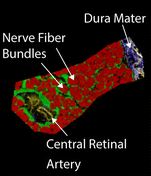
The human optic nerve carries signals from the retina to the visual cortex of the brain. Each optic nerve is comprised of approximately one million nerve fibers that are organized into bundles of 800-1200 fibers surrounded by connective tissue and supportive glial cells. Damage to the optic nerve contributes to a number of blinding diseases including: glaucoma, neuromyelitis optica, optic neuritis, and neurofibromatosis; however, the molecular mechanisms of optic nerve damage and death are incompletely understood. Herein we present high spatial resolution MALDI imaging mass spectrometry (IMS) analysis of lipids and proteins to define the molecular anatomy of the human optic nerve. The localization of a number of lipids was observed in discrete anatomical regions corresponding to myelinated and unmyelinated nerve regions as well as to supporting connective tissue, glial cells, and blood vessels. A protein fragment from vimentin, a known intermediate filament marker for astrocytes, was observed surrounding nerved fiber bundles in the lamina cribrosa region. S100B was also found in supporting glial cell regions in the prelaminar region, and the hemoglobin alpha subunit was observed in blood vessel areas. The molecular anatomy of the optic nerve defined by MALDI IMS provides a firm foundation to study biochemical changes in blinding human diseases.


No responses yet