Marcel Wiegelmann, Klaus Dreisewerd, Jens Soltwisch
J. J. Am. Soc. Mass Spectrom. (2016)
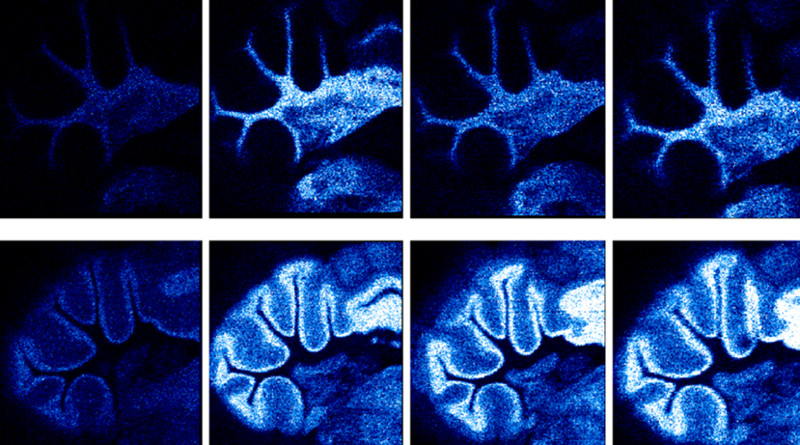
To improve the lateral resolution in matrix-assisted laser desorption/ionization mass spectrometry imaging (MALDI-MSI) beyond the dimensions of the focal laser spot oversampling techniques are employed. However, few data are available on the effect of the laser spot size and its focal beam profile on the ion signals recorded in oversampling mode. To investigate these dependencies, we produced 2 times six spots with dimensions between ~30 and 200 μm. By optional use of a fundamental beam shaper, square flat-top and Gaussian beam profiles were compared. MALDI-MSI data were collected using a fixed pixel size of 20 μm and both pixel-by-pixel and continuous raster oversampling modes on a QSTAR mass spectrometer. Coronal mouse brain sections coated with 2,5-dihydroxybenzoic acid matrix were used as primary test systems. Sizably higher phospholipid ion signals were produced with laser spots exceeding a dimension of ~100 μm, although the same amount of material was essentially ablated from the 20 μm-wide oversampling pixel at all spot size settings. Only on white matter areas of the brain these effects were less apparent to absent. Scanning electron microscopy images showed that these findings can presumably be attributed to different matrix morphologies depending on tissue type. We propose that a transition in the material ejection mechanisms from a molecular desorption at large to ablation at smaller spot sizes and a concomitant reduction in ion yields may be responsible for the observed spot size effects. The combined results indicate a complex interplay between tissue type, matrix crystallization, and laser-derived desorption/ablation and finally analyte ionization.


Comments are closed