 Young S. Park, Chong W. Yoo, Seok C. Lee, Sang J. Park, Jae H. Oh, Byong C. Yoo, Seung S. Paik, Kyeong G. Lee, So Y. Jin, Song C. Kim, Kwang P. Kim, Young H. Kim, Dongho Choi and Hark K. Kim
Clin. Chim. Acta. (2011), 412(21-22), 1978-1982
BACKGROUND: We evaluated whether direct tissue matrix-assisted laser desorption/ionization (MALDI) mass spectrometry (MS) analysis of lipids may distinguish intrahepatic cholangiocarcinomas from adjacent normal tissue and from other adenocarcinomas that frequently metastasize to liver.
METHODS: Four pairs of frozen surgical specimens of cholangiocarcinomas and adjacent normal tissue were analyzed using histology-directed, MALDI MS analysis. 2,5-dihydroxybenzoic acid / α-cyano-4-hydroxycinnamic acid were manually deposited on tumor-rich areas, and mass spectra were acquired using a MALDI-time of flight instrument.
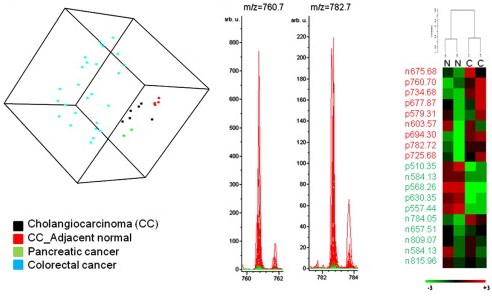
RESULTS: Cholangiocarcinomas and adjacent normal tissue samples demonstrated different lipid profiles, as evidenced by permutation P value<0.05 for the cross-validated misclassification rate. Cancer-associated lipid alteration was similar between cholangiocarcinomas and pancreatic cancers, but not between cholangiocarcinomas and colorectal cancers. Baseline lipid profiles were different between cholangiocarcinoma and colorectal cancers.
CONCLUSIONS: MALDI MS analysis of lipid distinguishes cancerous epithelium of cholangiocarcinoma from adjacent normal tissue, and between cholangiocarcinomas and colorectal cancers.
No responses yet