 Kevin R. Tucker, Eric J. Lanni, Leonid A. Serebryannyy, Stanislav S. Rubakhin and Jonathan V. Sweedler
Anal. Chem. (2011), 83 (23), 9181-9815
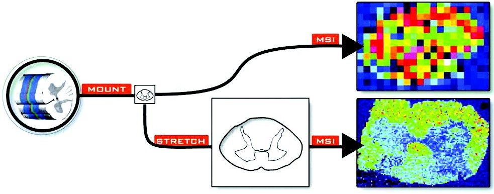
Matrix-assisted laser desorption/ionization (MALDI) mass spectrometry imaging (MSI) combines information-rich chemical detection with spatial localization of analytes. For a given instrumental platform and analyte class, the data acquired can represent a compromise between analyte extraction and spatial information. Here, we introduce an improvement to the spatial resolution achievable with MALDI MSI conducted with standard mass spectrometric systems that also reduces analyte migration during matrix application. Tissue is placed directly on a stretchable membrane that, when stretched, fragments the tissue into micrometer-sized pieces. Scanning electron microscopy analysis shows that this process produces fairly homogeneous distributions of small tissue fragments separated and surrounded by areas of hydrophobic membrane surface. MALDI matrix is then applied by either a robotic microspotter or an artist’s airbrush. Rat spinal cord samples imaged with an instrumental resolution of 50–250 μm demonstrate lipid distributions with a 5-fold high spatial resolution (a 25-fold increase in pixel density) after stretching compared to tissues that were not stretched.
No responses yet